Biomolecular Condensates in Biology & Disease
University
St. Jude
Presenter
Dr. Richard Kriwacki

During his >25 year career, Richard Kriwacki has sought to understand the molecular basis of regulation of essential biological processes (e.g., cell division, apoptosis, ribosome biogenesis, and transcription) with emphasis on the roles of intrinsically disordered proteins (IDPs) and phase separation in these processes. The lab applies structural biology and biophysical techniques (NMR spectroscopy, x-ray crystallography, calorimetry, AUC, etc.), as well as biochemical, cell biological and cell imaging methods, to study the details of biomolecular mechanisms from the test tube to cells.
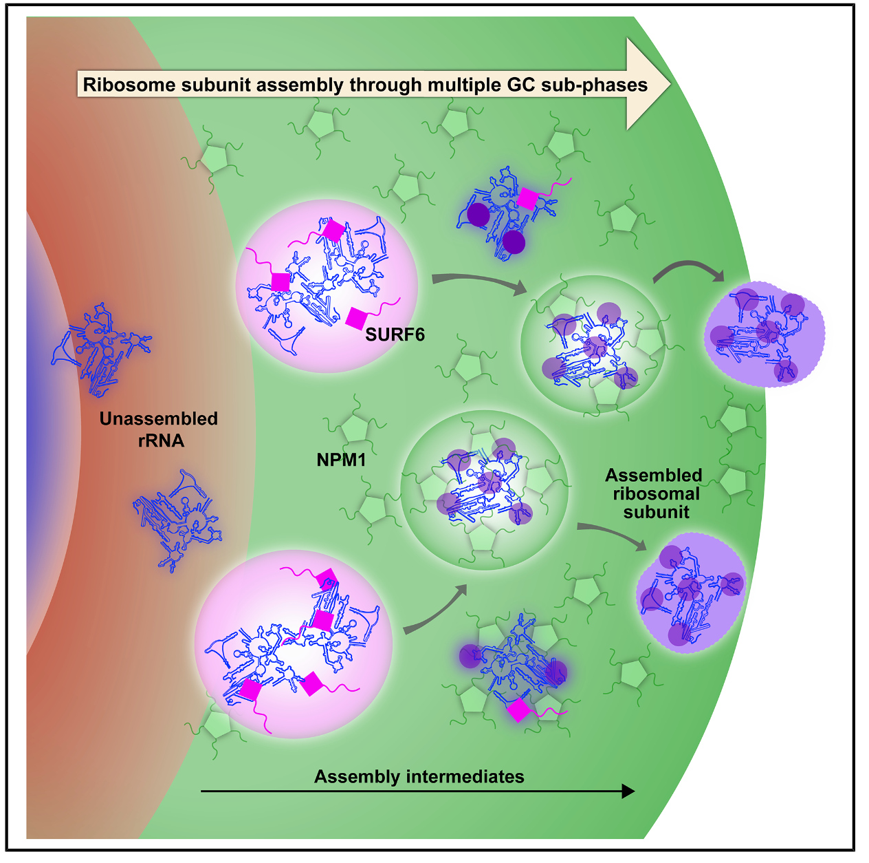
One research direction focuses on the role of phase separation in biology & disease, specifically the role of Nucleophosmin (NPM1) and other proteins in the structure, dynamics and function of the liquid-like membrane-less organelle, the nucleolus. A primary goal is to understand how phase separation contributes to the molecular steps underlying ribosome biogenesis.
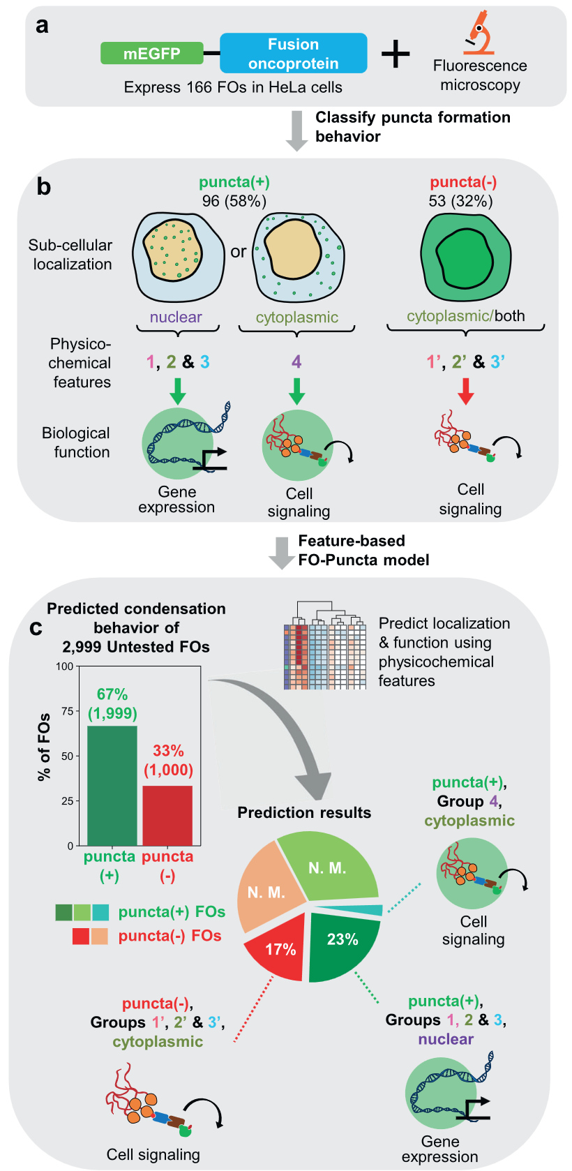
Another research direction addresses the role of phase separation by fusion oncoproteins (FOs) in cancer. We discovered that some FOs would undergo phase separation (PS) in the nuclei of cells, forming aberrant transcription centers that drive aberrant gene expression and transform cells. Broadly, we seek to understand the prevalence of phase separation by FO across diverse pediatric cancers and to apply this knowledge to advance cancer therapeutics.